
 News Trial Stats
Mon–Thu 8am–4:30pm CT · Fri 8am–3pm CT
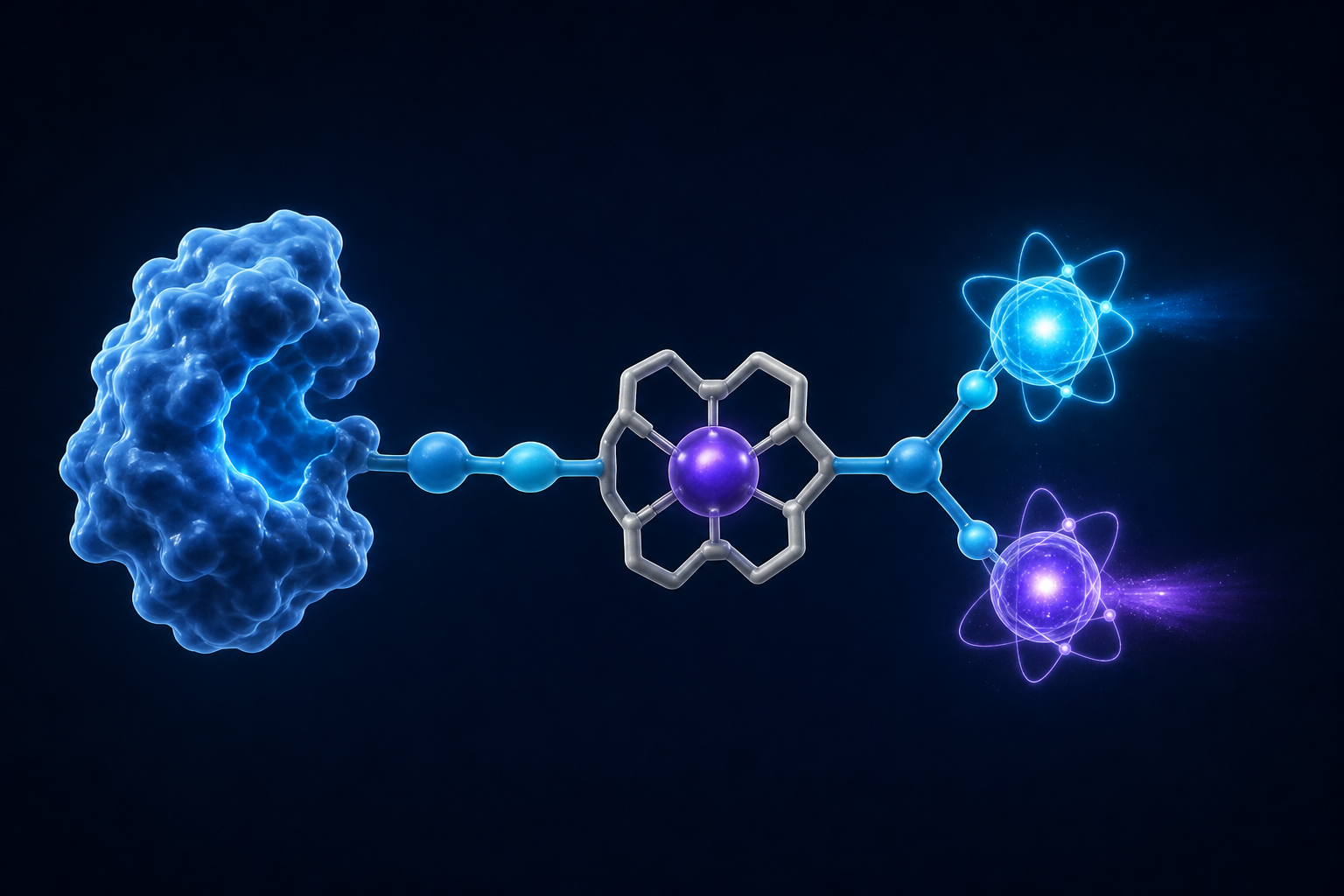
Parts of a Theranostic Drug — 01 · The Target
Radioligand Targets
The marker on a cancer cell that the entire drug is built to find.
Every other part of a radiopharmaceutical is manufactured and injected. The target isn’t — it’s already in the body, on the cancer itself. Understand the target and you understand the whole treatment.
Start here
What a target is
Target
A specific identifier — a protein, glycoprotein, or carbohydrate — found on a cancer or group of cancers. This identifier becomes the imaging and therapeutic target for the radiopharmaceutical’s ligand.
The one thing to understand first
The target is part of the cancer, not part of the drug
The ligand, linker, chelator, and isotope are assembled into a single molecule and injected. The target is a structure — almost always a protein — that already sits on the surface of the cancer cells. The drug is built to fit a lock that is already there.
This is why the target comes first. Before anyone designs a ligand, chooses a chelator, or picks an isotope, they answer one question: what does this cancer display that healthy tissue does not? That answer is the target, and every other part is engineered around it.
It is also the single biggest factor in whether a treatment can help — and one you can check in advance, because the same target that therapy binds is the one an imaging scan lights up. If the drug is a key, the target is the lock, installed on the cancer before the key is ever made.
Why it decides everything
Four things the target controls
Whether treatment is possible
No target, no therapy. The tumor must display the marker the drug is built for — and a diagnostic scan can confirm that before treatment begins.
How well it works
The more strongly the cancer displays the target, and the more of its cells that do, the more drug binds and the higher the dose delivered to the tumor.
What side effects to expect
Wherever the same target appears on healthy tissue, that tissue gets some radiation too — so effects are predictable in advance from biology.
Whether the cancer escapes
If tumor cells stop displaying the target, the drug loses its grip. The cells that survive are often the ones that showed the least target to begin with.
What makes a good one
The ideal target
Finding a good target is the hardest part of building one of these drugs. The ideal has several properties at once, and real targets are always a compromise among them.
-
Strongly present on the cancer. More target means more binding sites — more radiation delivered where it is wanted.
-
Absent — or nearly so — on healthy tissue. The property that sets the safety margin. Shared with a vital organ, it becomes a problem.
-
On most of the tumor’s cells, not just some. If only a fraction display it, the rest go untreated and seed a relapse.
-
Stable, not easily switched off. Cancers that can stop producing a target under pressure will do exactly that.
-
Reachable on the cell surface. A circulating drug has to physically get to it; a marker locked inside the cell is out of reach.
-
Bonus: it pulls the drug inside. Some targets draw the drug into the cell and trap it, keeping radiation near the nucleus.
The patient payoff
Side effects are written into the target
In most cancer treatments, side effects are hard to predict. In radioligand therapy they often aren’t — they follow directly from where the target also appears on normal tissue.
| If the target is… | The common effect is | Because |
|---|---|---|
| PSMA | Dry mouth, dry eyes | Salivary and tear glands also carry PSMA |
| Somatostatin receptor | Nausea, hormonal shifts | Present in the GI tract and pituitary |
| NIS (iodine) | Salivary changes; thyroid uptake | Salivary glands share the transporter; thyroid uptake is intended |
| CD20 | Temporary drop in immune B-cells | Healthy B-cells share the marker, then recover |
This is why your care team’s precautions are specific, not generic. They know the target, so they know which tissues to watch — and can often tell you what to expect before it happens.
Wherever a target also appears on healthy tissue, that tissue receives some radiation too — so a drug’s likely side effects can often be predicted in advance from the biology of its target. That predictability is part of what makes theranostics distinctive.
The core limitation
One target is a single point of failure
Today’s approved drugs are built around a single target. That keeps them simple, but it creates two problems that recur across the field.
The target is on healthy tissue too
No target is found only on cancer. Wherever it also appears on normal tissue, that tissue receives radiation — which sets a ceiling on how much drug can safely be given.
The cancer can drop the target
Under treatment pressure, tumor cells that stop displaying the target survive and multiply. The therapy stops working — not because the drug failed, but because the target disappeared.
Both trace back to the same root: one drug, one target. That is the ceiling the next generation of designs is built to break.
What’s coming
Beyond one target at a time
Two targets at once
Bispecific designs stick only to cells displaying both of two markers — a combination healthy tissue rarely has. Sharper aim, wider safety margin.
Pan-cancer targets
FAP sits in the supportive tissue around many tumor types rather than one organ, raising the prospect of one approach that works across cancers.
Combination targeting
Hitting two markers in parallel leaves fewer escape routes — dropping one target is no longer enough to evade treatment.
New target discovery
The field’s bottleneck is finding markers strong on cancer and quiet on healthy tissue. Much of current research is that search.
Explore Targets
Find a target by cancer
Every marker currently being imaged or treated in radioligand therapy. Choose a cancer, or search by name.
All targets
Loading targets…
Share your feedback
Help us improve your experience