
 News Trial Stats
Mon–Thu 8am–4:30pm CT · Fri 8am–3pm CT
Patient Education — Interpreting Your Scans
How to Interpret Your Scans
Tape measures and light bulbs — what CT, MRI and PET can and can’t tell you about cancer.
CT and MRI measure how big something is. PET shows whether it carries a target — and, by how brightly it lights up, how likely it is to be cancer. Knowing the difference makes your scan report far less mysterious.
Start here
Every scan answers a different question
Scan reports are written for doctors, and words like “lesion,” “enhancing,” or “avid” can make any patient anxious. The single most helpful thing to know is that different scans answer different questions. Some show the body’s structure. Others show what cells are actually doing.
CT & MRI
Anatomic imagingThe tape measures
“Where is it, and how big is it?”
They create detailed pictures of shape, size and location — excellent for measuring a mass and seeing whether it grows or shrinks over time.
PET Scan
Molecular imagingThe light bulbs
“Does it carry the target — and how likely is it to be cancer?”
A tracer travels to a specific target and lights up wherever it finds it. How bright it glows (its SUV) compared with normal tissue such as the liver helps show whether a suspicious spot is likely cancer.
“Lesion”
A neutral word for any area that looks different from the tissue around it. It does not mean cancer. Cysts, scars, benign growths and inflammation are all “lesions” too.
Great at size, not at identity
Think of a CT or MRI as a very precise tape measure. It can tell your doctor that there is a 2-centimeter mass in the liver, exactly where it sits, and what it’s next to. What it usually can’t tell is what that mass actually is.
A benign cyst, an old scar, a patch of infection, a harmless growth and a tumor can all look like a spot of similar size and shape. Radiologists can often say whether something looks suspicious or reassuring, but a picture of size and shape alone rarely proves cancer.
That proof comes from a biopsy — a small sample of tissue looked at under a microscope by a pathologist. Once a biopsy confirms a mass is cancer, the tape measure becomes one of the most important tools in your care.
How CT and MRI fit into your cancer journey
- 1
Something is found
A CT or MRI shows a mass or spot. At this point it is only a size and a location — not yet a diagnosis.
- 2
A biopsy answers “what is it?”
Tissue is sampled and examined. This is what confirms cancer, its type, and often the markers that guide treatment.
- 3
A baseline is measured
Before treatment starts, the known tumors are measured carefully. This becomes the starting point every later scan is compared against.
- 4
Follow-up scans track response
Repeat scans re-measure the same tumors to see whether they are shrinking, stable, or growing — and whether any new spots have appeared.
Reading the tape measure over time
In clinical trials and many practices, response is measured with a standard rulebook (often called RECIST) that adds up the longest diameters of selected tumors and compares them with the baseline.
Shrinking
Response
Measured tumors have shrunk meaningfully — under RECIST, at least about 30% smaller in total. Complete response means the measured tumors are no longer visible.
Holding
Stable disease
Not shrinking enough to count as response, not growing enough to count as progression. For many treatments, stable is a good result.
Growing
Progression
Measured tumors have grown meaningfully — under RECIST, about 20% larger than their smallest point — or a clearly new tumor has appeared.

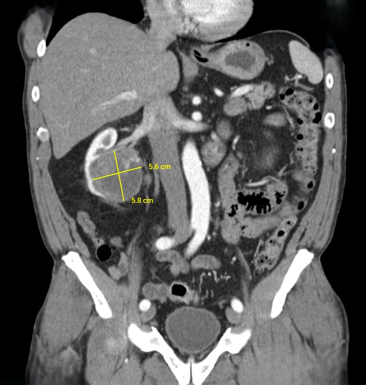
The same mass measured on two CT scans. Comparing its size against the baseline is how your team judges whether treatment is working.
Size can lag behind biology. A tumor that has been successfully treated may leave scar tissue that keeps the same size for months, and some treatments like immunotherapy can briefly make tumors look bigger from inflammation before they shrink. That’s why one scan is rarely read alone — your team looks at the trend.
Under the hood
How CT and MRI work — and where each shines
Both create cross-sectional “slices” of the body, but they get there in very different ways. That difference decides which parts of the body each one sees best.
Fast X-ray slices
A CT scanner rotates an X-ray beam around you and a computer stacks the images into detailed slices. It uses ionizing radiation — a CT of the chest, abdomen and pelvis delivers roughly the same dose as a few years of natural background radiation. An iodine contrast dye is often given through an IV to make blood vessels and organs stand out.
- Very fast — usually seconds to a few minutes
- Freezes motion, so it handles breathing well
- The workhorse for lungs, chest, abdomen and pelvis
- Widely available, good for whole-body staging
- Uses radiation, which adds up over many scans
- Less detail in soft tissues like brain and prostate
- Contrast dye can be a concern with kidney problems or allergy
Magnets and radio waves
An MRI uses a very strong magnet and radio waves to read signals from the water in your tissues. There is no ionizing radiation. Scans take longer — often 30 to 60 minutes — and you need to hold very still, because movement blurs the images. A gadolinium contrast dye is sometimes used.
- No radiation
- Outstanding soft-tissue detail
- Best for still, solid structures: brain, spine, bone and joints, prostate, liver
- Motion blurs images — poor for lungs, which move with every breath
- Long, loud and enclosed; can be hard with claustrophobia
- Some metal implants and devices need screening first
CT is usually preferred for
MRI is usually preferred for
Seeing the target, not just the shape
A PET scan works on a completely different idea. You receive a small injection of a tracer — a diagnostic radioisotope attached to a ligand that seeks out one specific target. Remember the truck from How Theranostics Work: for a PET scan, the cargo is light bulbs. Wherever the truck finds its target, the bulbs switch on, and the scanner records the glow.
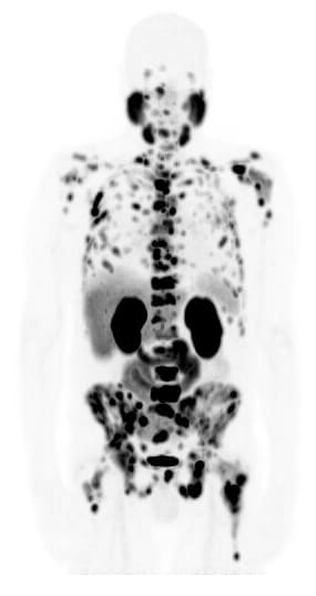
PET images are almost always fused with a CT or MRI taken at the same visit (PET/CT or PET/MRI). The CT or MRI provides the map; the PET shows which spots on that map are lit up. Brightness tells you about the biology — including how likely something is to be cancer — by showing how much of the target is there or how active the cells are, rather than just size.
When a suspicious spot lights up, it tells you two things: the target is there, and — depending on how bright it is (its SUV) — whether it is likely to be cancer. A spot glowing far brighter than normal tissue such as the liver is much more likely to be cancer; one glowing at or below liver level is less likely to be.
Because each tracer looks for a different target, the type of tracer matters enormously. A spot that lights up on one tracer may be dark on another.

| Tracer “light bulb” (isotope) | What it seeks | Example cancers |
|---|---|---|
| Non-specific — activity, not a single target | ||
F-18 | FDG — sugar uptake Busy, fast-growing cells burn more glucose. Lights up many cancers, but also inflammation and infection. Approved | Lung, lymphoma, melanoma, head & neck, colorectal, esophageal and many more |
| Target-specific — seeks one marker on the cancer | ||
Ga-68F-18Cu-64 | PSMA Prostate-specific membrane antigen, on most prostate cancer cells. Approved | Prostate cancer |
Ga-68Cu-64 | SSTR (somatostatin receptor) DOTATATE / DOTATOC tracers; companion scan for Lutathera. Approved | Neuroendocrine tumors, some lung carcinoids, pheochromocytoma |
Ga-68F-18 | FAP Fibroblast activation protein, on the support tissue surrounding many tumors (FAPI tracers). Investigational | Pancreatic, sarcoma, breast, lung, colorectal and many other solid tumors |
Ga-68F-18 | ACP3 Prostatic acid phosphatase, a newer prostate cancer target. Investigational | Prostate cancer |
Zr-89Ga-68 | STEAP1 A surface protein highly expressed on prostate cancer. Investigational | Prostate cancer; also studied in Ewing sarcoma |
Zr-89Ga-68 | DLL3 Delta-like ligand 3, on neuroendocrine-type cancer cells. Investigational | Small cell lung cancer, neuroendocrine prostate cancer |
Zr-89Ga-68 | B7-H3 An immune-checkpoint protein found on many solid tumors. Investigational | Prostate, lung, neuroblastoma, pediatric brain tumors and others |
Isotopes shown are common examples, not a complete list. Zr-89 is typically paired with antibody-based tracers. Investigational tracers are available through clinical trials.
In theranostics, the PET scan does double duty: if the target lights up on the scan, the matching therapy has somewhere to go. That is why a PSMA or DOTATATE PET is often required before radioligand therapy.
SUV: how bright is the light bulb?
PET reports often list a number next to a spot, such as “SUVmax 8.4.” SUV stands for Standardized Uptake Value — think of it as the spot’s brightness score. It measures how much tracer collected in that spot compared with what you’d expect if the tracer had spread evenly through your whole body.
In general, the brighter a suspicious spot, the more likely it is to be cancer. But the number is most meaningful when it’s compared with something. Reports usually also list a reference score from normal, non-cancerous tissue — most often the liver — so you can see how your spot measures up. A spot that scores lower than the reference is unlikely to be cancer; one that scores well above it is much more likely to be.
SUV = 1
The tracer is no more concentrated there than average — the same as if it had spread evenly everywhere. An SUV of 6 means about six times the average concentration. SUVmax is the single brightest point within a spot, and it is the value most reports use.
The liver: your built-in reference point
An SUV number means little on its own. What matters is how it compares with normal tissue on the same scan — most often the liver, which takes up a steady, predictable amount of most tracers (the blood pool and salivary glands are also used). Comparing a suspicious spot with the liver turns a number into a useful judgment:
Less likely cancer
A spot that glows less than normal liver is less likely to be cancer — often inflammation, healing, normal tissue, or something too small to call. It is frequently watched on the next scan.
Indeterminate
A spot about as bright as liver is a gray zone. Location, size on CT or MRI, and change since the last scan help decide — and sometimes a biopsy or repeat scan is recommended.
Likely cancer
A spot glowing significantly brighter than liver — especially in a place and pattern that fits your known cancer — is likely cancer. For radioligand therapy, uptake above liver also suggests the treatment has a strong target.
An illustrative brightness scale (FDG example)
Roughly the level of blood in the large vessels — often around 1.5–2.
Similar to or a little below normal liver — often around 2–3. Frequently inflammation, healing or normal tissue.
Clearly brighter than liver. More suspicious, especially in a pattern that fits the known cancer.
Many times brighter than background. Often seen in active cancer — though infections can be bright too.
There is no single “cancer cutoff.” Normal ranges differ by tracer, scanner, timing, blood sugar and body size. That’s why radiologists usually compare a spot with reference organs on the same scan — such as the blood pool, liver, or salivary glands — rather than relying on one fixed number.
What SUV is most useful for
Pattern
Context, not a verdict
A bright spot in a place that fits your known cancer means something different than a faint spot in a place that commonly glows for harmless reasons.
Over time
Spotting new areas
On follow-up PET scans, whether new spots have lit up since the last scan is often more telling than small changes in the brightness of a spot you already had.
Therapy
Selecting treatment
For radioligand therapy, strong uptake (often brighter than liver) suggests the target is plentiful enough for the treatment to deliver its dose.
Very important
A change in SUV is not a proven measure of whether treatment is working. If the same spot scores higher or lower on a later scan, that alone has not been validated to mean the cancer is growing or shrinking — especially for the targeted PET scans used in theranostics, such as PSMA and somatostatin-receptor scans.
SUV can shift for many reasons unrelated to the cancer: a different scanner, a different tracer or dose, the time between injection and scanning, or treatment changing how much target the cells display. Response is judged on the whole picture — size on CT or MRI, new spots, blood markers like PSA, and how you feel — not on one spot’s brightness score.
An important caution
Not everything that lights up is cancer
A PET tracer goes wherever its target is — and many targets exist in healthy tissue too. Faint, low-SUV spots are especially common and are very often harmless. Here are the usual reasons something glows without being cancer.
Normal organs
Every tracer has places it naturally collects or leaves the body through. FDG lights up the brain and heart; PSMA tracers light up the salivary glands and kidneys; DOTATATE lights up the spleen. These are expected and are not disease.
Inflammation, infection & healing
Immune cells are hungry for sugar, so FDG often glows in infections, pneumonia, arthritis, recent surgery, biopsy sites, radiation changes, and lymph nodes reacting to a recent vaccine.
PSMA tracers can mildly light up healing fractures, nerve clusters called ganglia, and some benign bone conditions.
Low SUV, low concern
A spot with low uptake — near background or below liver — and no matching change on CT or MRI is frequently benign or simply too small to call. Reports may say “nonspecific” or “likely inflammatory.”
These are often watched on the next scan rather than treated.
The other direction
And not every cancer lights up brightly. Some cancers use little sugar, so they look faint on FDG — including many prostate cancers, some low-grade or slow-growing tumors, and certain breast and neuroendocrine cancers. Very small spots may also be below what PET can detect.
This is exactly why choosing the right tracer for the right cancer matters, and why PET findings are always read together with CT or MRI, your history, and — when needed — a biopsy.
A glow is a clue, not a diagnosis. Brightness, location, pattern, how it compares with the last scan, and what CT or MRI shows all have to fit together. If a finding truly matters to your treatment, your team may recommend a biopsy to be sure.
Putting it to use
Questions to Ask Your Physician about Your Reports
It’s normal to read your report in a patient portal before you’ve spoken to your doctor — and normal to feel worried by it. These questions can turn a confusing report into a useful conversation.
-
Is this spot new, or was it there before? Comparison with earlier scans is often the most informative part of a report.
-
Has the cancer been confirmed by biopsy? If not, a CT or MRI finding alone may not be enough to know what it is.
-
Overall, is it growing, shrinking, or stable? Ask about the trend across scans, not just a single measurement.
-
Which PET tracer was used, and what was the SUV compared with liver or background? That context explains whether a glow is concerning or likely harmless.
-
Could this be inflammation, infection, or healing? Mention any recent surgery, injury, infection or vaccine.
-
Does this change my treatment — or open up a trial? A positive PSMA or DOTATATE scan, for example, may make you eligible for radioligand therapy or a theranostic clinical trial.
CT and MRI tell you how big. PET tells you what it’s doing. A biopsy tells you what it is. Your care team puts all three together — so bring your questions, and never let a single number or word on a report decide how you feel before that conversation.
Keep going
Where to next
Educational content, not medical advice. Scan findings must be interpreted by your radiologist and care team in the context of your full history. Always discuss your specific results with your Theranostics care team.
Share your feedback
Help us improve your experience